On September 26, 2024, over 30 participants were able to listen to a great lecture delivered by Dr. Pradesh Ghimire, who talked about the role of imaging in acute stroke care. The presentation underscored the critical importance of CT and MRI imaging in stroke diagnosis.
Dr. Ghimire emphasized that the most essential imaging in stroke care revolves around CT and how to use it to differentiate between ischemic and hemorrhagic strokes. He introduced the “2P” approach – focusing on the parenchyma (brain tissue) and the pipes (vascular structures), as well as the “3C” approach, which includes imaging for collaterals, the core (including the penumbra), and clots. This ensures that both the brain tissue and blood vessels are comprehensively evaluated, before adding additional imaging techniques to get a more in-depth assessment of stroke severity and progression.
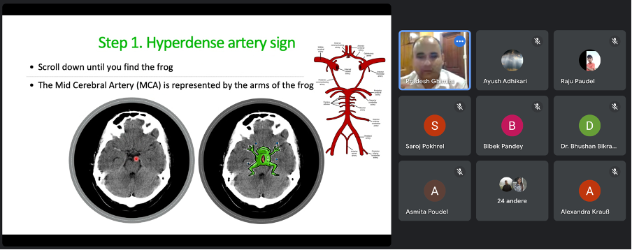
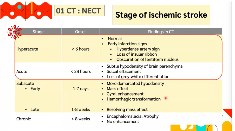
Depending on the stage of the stroke, different findings will appear on a CT scan. For ischemic strokes, early signs like the hyperdense artery sign can be critical for early diagnosis. Dr. Ghimire also discussed the use of the ASPECTS (Alberta Stroke Program Early CT Score) to quantify early ischemic changes on CT brain scans in acute stroke patients, which helps assess the extent of early infarction.
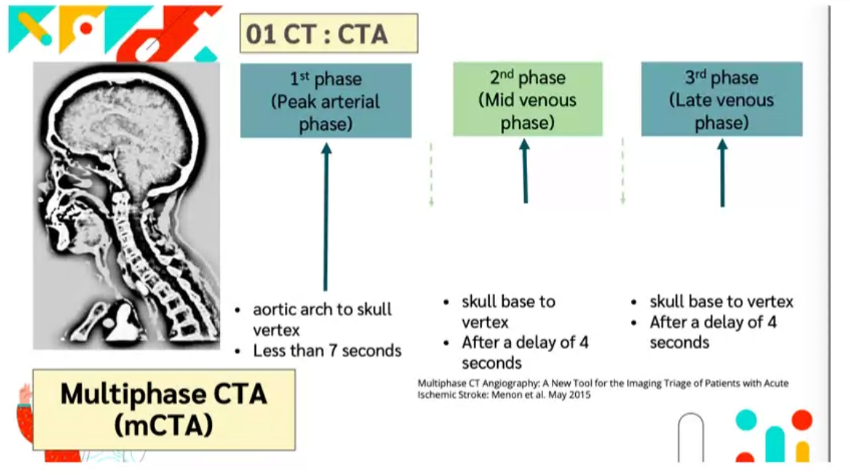
While Non-Contrast Enhanced CT (NCT) is commonly used, it has certain limitations, particularly its low sensitivity for detecting small or subcortical infarcts. To address this, CT Angiography (CTA) is utilized due to its high sensitivity and specificity in evaluating brain vessels and collaterals. Additionally, CT Perfusion imaging is valuable for assessing the brain’s microcirculation and the state of the stroke penumbra, which is essential in determining tissue viability.
In addition to CT scans, MRI can be used for further evaluation, with sequences such as Diffusion-Weighted Imaging (DWI), Apparent Diffusion Coefficient (ADC), Fluid-Attenuated Inversion Recovery (FLAIR), and T2-FLAIR being particularly useful. Furthermore, Time-of-Flight (TOF) imaging, which does not require contrast administration, is effective in visualizing blood flow within vessels.
Dr. Ghimire concluded by reiterating the pivotal role of imaging not only in initial stroke diagnosis but also in the evaluation of wake-up strokes and determining the appropriateness of intravenous thrombolysis (IV rtPA) and endovascular treatment. The session ended with a lively Q&A segment, where Dr. Ghimire addressed numerous questions from the audience. Join us again for the next lecture on October 17, 2024. Jessica Golenia will discuss “Hyperacute Plus Acute Stroke Care – Nurse Tasks.”
Plese send me the free “Stroke Knowledge” materials: